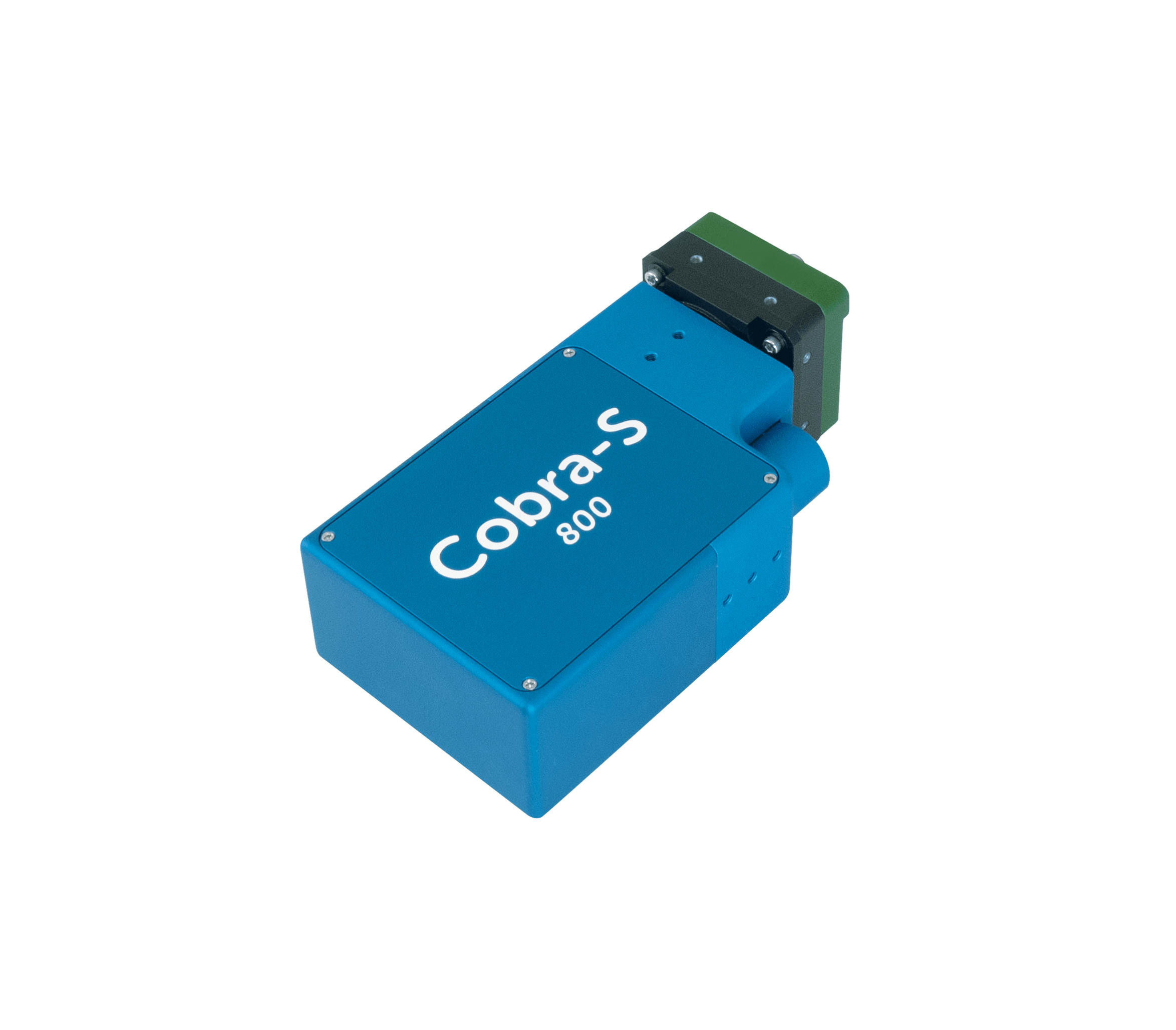
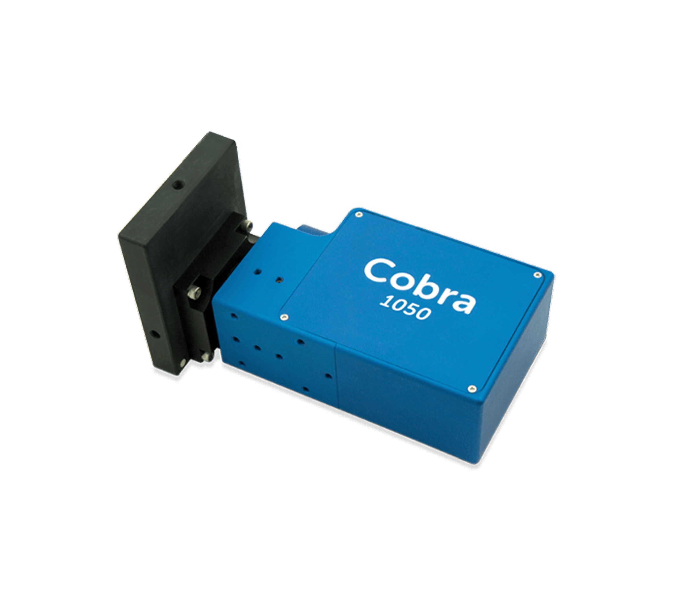
Cobra series spectrometers are at the leading edge of eye research
Modern ophthalmic SD-OCT systems must image through scattering tissue at high speed and high sensitivity, placing stringent constraints on spectrometer design. An OCT spectrometer must maximize optical throughput, maintain accurate dispersion for precise Fourier reconstruction, and provide polarization-independent efficiency, while supporting high acquisition rates for real-time retinal imaging without compromising signal integrity.
Spectrometers must maintain stable performance across variations in temperature, humidity, and pressure, withstand shock and vibration during transport, installation, and use, and remain compatible with clinical electromagnetic environments. Long-term alignment stability and manufacturing repeatability are essential to ensure reliable operation over the device lifetime.
As a result, OCT spectrometer design has evolved from general laboratory instrumentation into highly specialized subsystems engineered for medical imaging applications.
Features & Benefits
•
Exceptional 1st order diffraction efficiency gratings
•
Polarization independent efficiency across the full spectral band
•
Exceptional roll-off performance
•
High speed line cameras to avoid fringe washout
•
Long term stability and manufacturing reproducibility
•
Robust design & SDKs for ease of integration
We offer high-efficiencient, robust, and integration-ready OCT spectrometer components for superior imaging performance.
OCT Imaging in Action
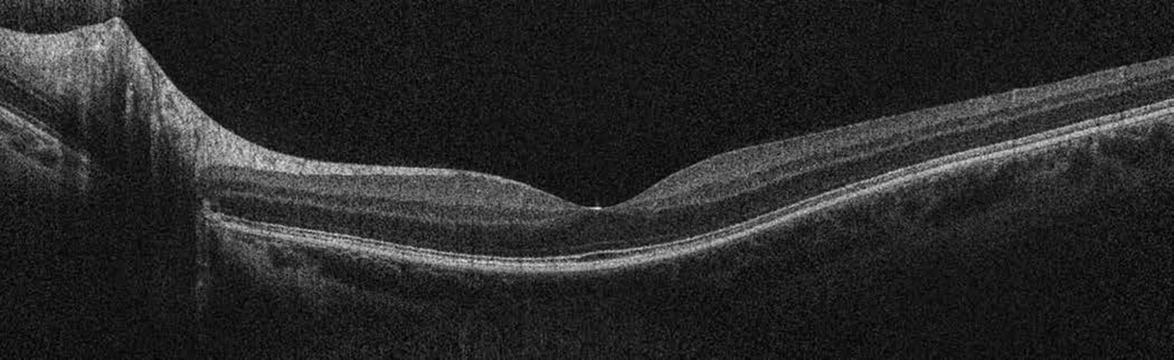
Retina Scan
An ultrafast SD-OCT image of human retina provides both wide-field and high-resolution images. This image was acquired using a Cobra-S OCT spectrometer. No averaging of the data was performed. The image was acquired at a 128-kHz A-line rate, with an calculated axial resolution of 3 μm in tissue.
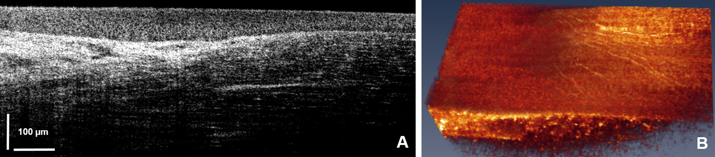
Cornea Scan
These high-resolution corneal images were taken with a system built using a Cobra-S 800 spectrometer: (a) A cross section of a healthy cornea showing normal tear film (TF), (b) In vivo volume of the healthy cornea, (c) Similar layers in a keratoconus patient showing irregular TF and loss of structure between epithelium and Bowman’s layer, and (d) In vivo volume of the keratoconus eye. EPI: epithelial layer; BM: Bowman’s layer; STR: stroma.
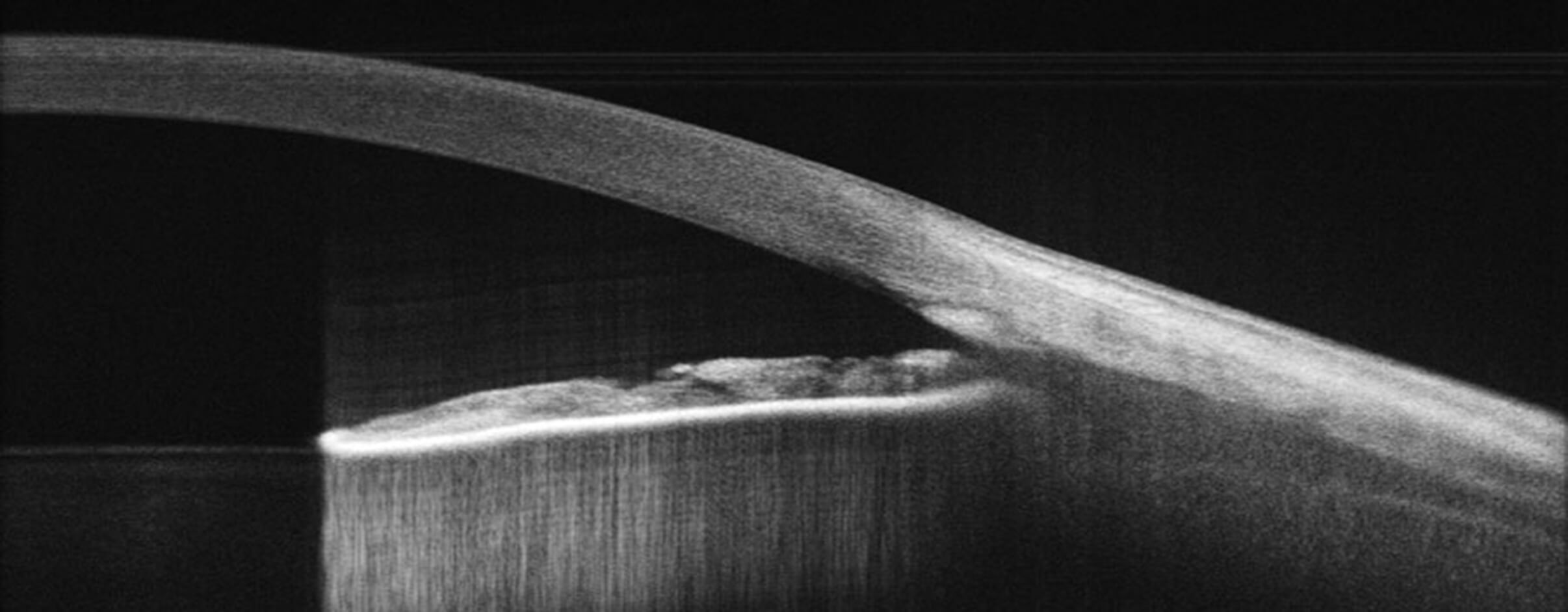
Anterior Segment
Cross-section of anterior segment of human eye showing iridocorneal angle, the angle formed between the cornea and the iris, taken using a Cobra 1300 OCT spectrometer. The structure is considered important in diagnosing eye conditions like glaucoma.
Misty Johnson is responsible for taking care of our grating customers at the Logan, Utah, facility. She has been a member of the Wasatch Photonics team since 2009. By understanding her customers’ needs and goals, as well as the current grating market, Misty is able to provide a superior level of customer support. As Account Manager, she acts as a liaison between customers and Wasatch Photonics’ manufacturing department making sure that the lines of communication are always open.
Customer support is an integral part of Wasatch Photonics commitment to customers. Whether you are in the market to purchase a single grating, inquiring as a distributor, or purchasing large volume, Misty will guide you seamlessly through the custom design process. Misty strives to build trust and strong long term relationships with Wasatch Photonics customers. She brings 20 years’ experience as customer support manager from a number of manufacturing and service companies.