OCT-A represents a natural evolution of OCT, extending structural imaging into functional vascular mapping. By detecting motion contrast from red blood cells, it enables dye-free, depth-resolved visualization of microvasculature with unmatched detail compared to other imaging modalities.
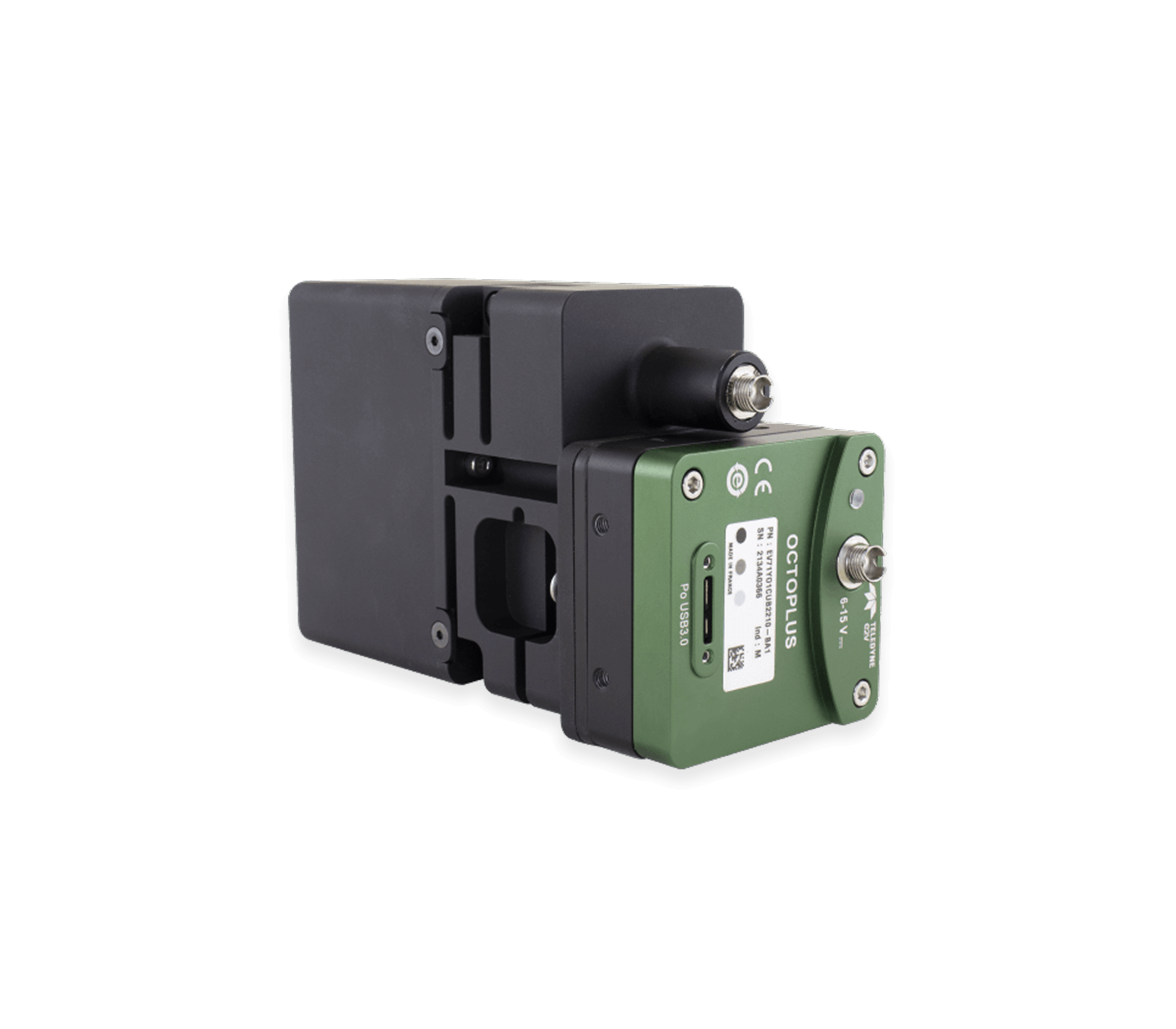
This capability critically depends on system stability, acquisition speed, and sensitivity. For OCT system developers, spectrometer design becomes even more specialized in OCT-A applications, where high optical throughput, fast detection, and long-term stability are essential for reliable angiographic performance.
As OCT-A expands across ophthalmology, oncology, and dermatology, advanced spectrometer technology remains central to unlocking its clinical and research potential. Wasatch continues to lead in enabling these developments.
Features & Benefits
•
High optical throughput spectrometer
•
High speed acquisition cameras
•
Spectrometer stability (thermal and mechanical) is a must
•
OCT-A maps blood flow without the need for injectable dyes
•
Provides 3-D resolved images vs 2-D of traditional angiography
•
Benefits from high axial resolution
Angiography at Wasatch is a word for high-speed, high-resolution OCT-A imaging with stable, dye-free 3D flow mapping.
Angiography in Action
OCT-A Dermatology
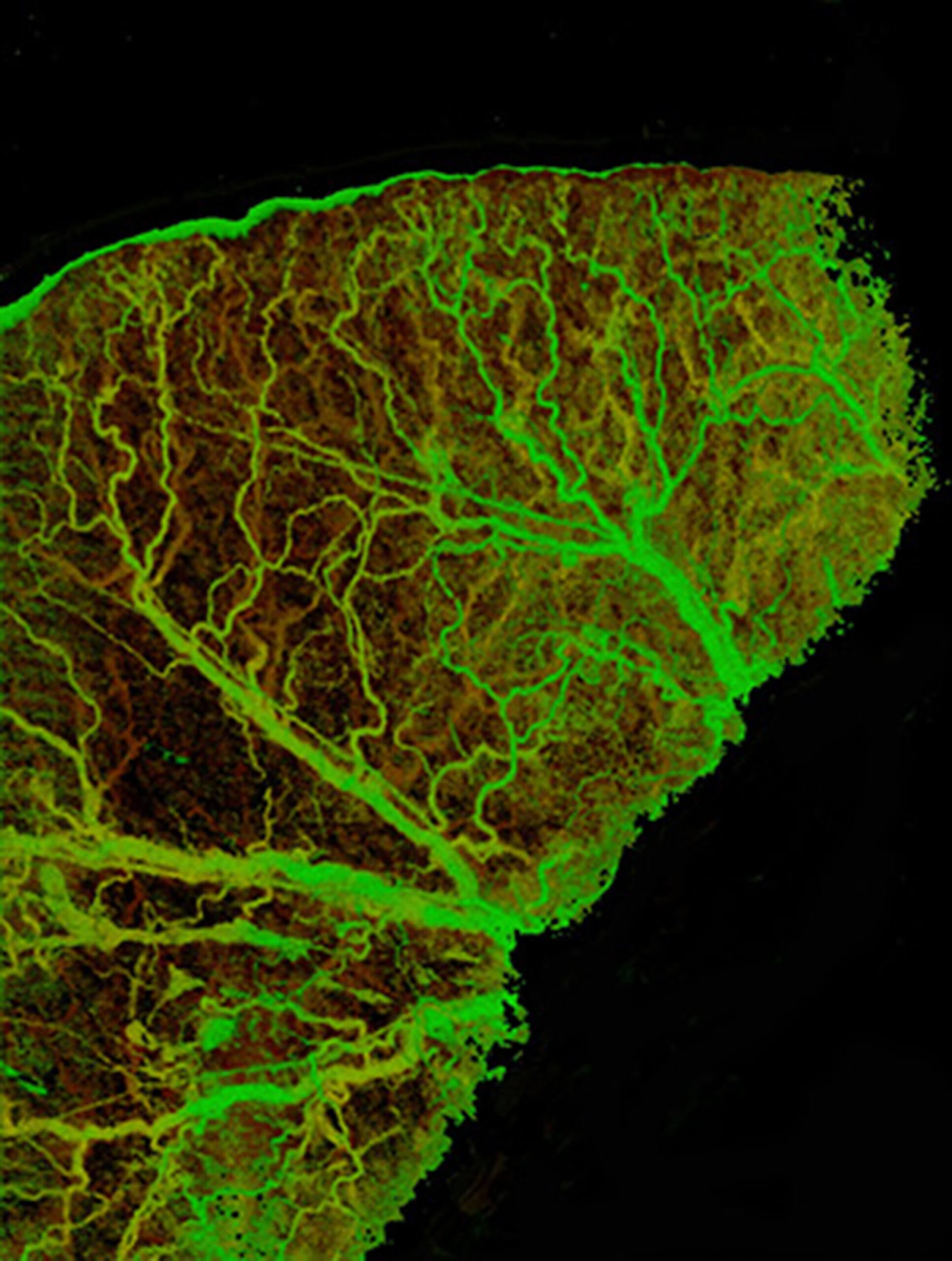
OCT angiography (OCTA) image of a full mouse ear taken with a Cobra 800 OCT spectrometer, showing a well-defined vascular pattern. This image demonstrates the high resolution that can be achieved with OCT angiography, which translates well to imaging vasculature of the skin.
OCT-A Oncology
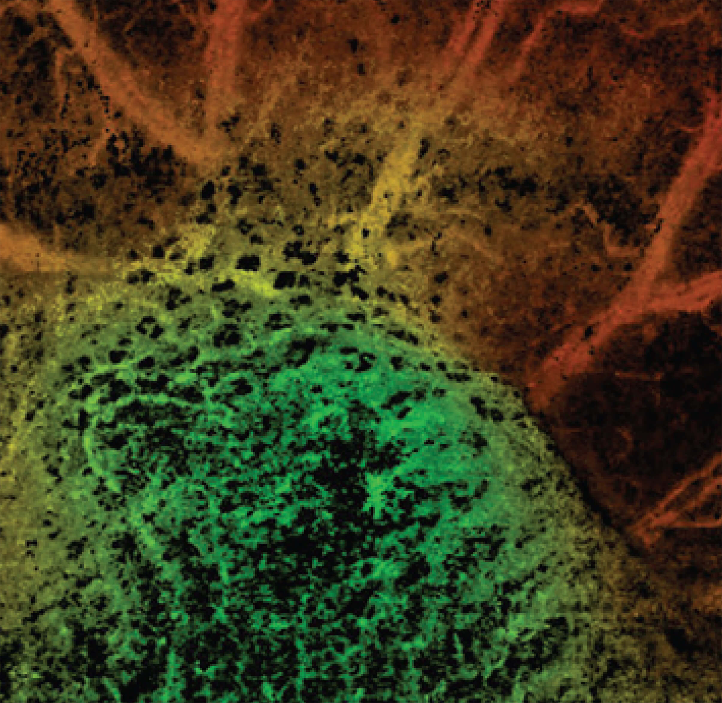
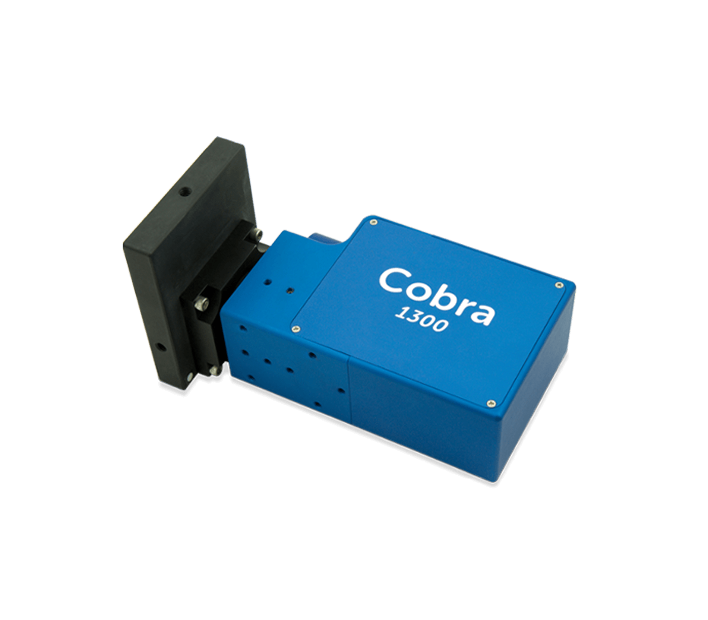
OCT angiography (OCTA) image of a diseased mouse ear, taken with a Cobra 1300 OCT spectrometer. In this case, a tumor was implanted in the mouse ear to demonstrate the ability of OCT angiography to distinguish tumor margins. This technique is of particular interest in cancer and carcinoma treatment. It can also be applied in endocrinological diseases like diabetes, in which there is a need to study perfusion in the body.
OCT-A Ophthalmology
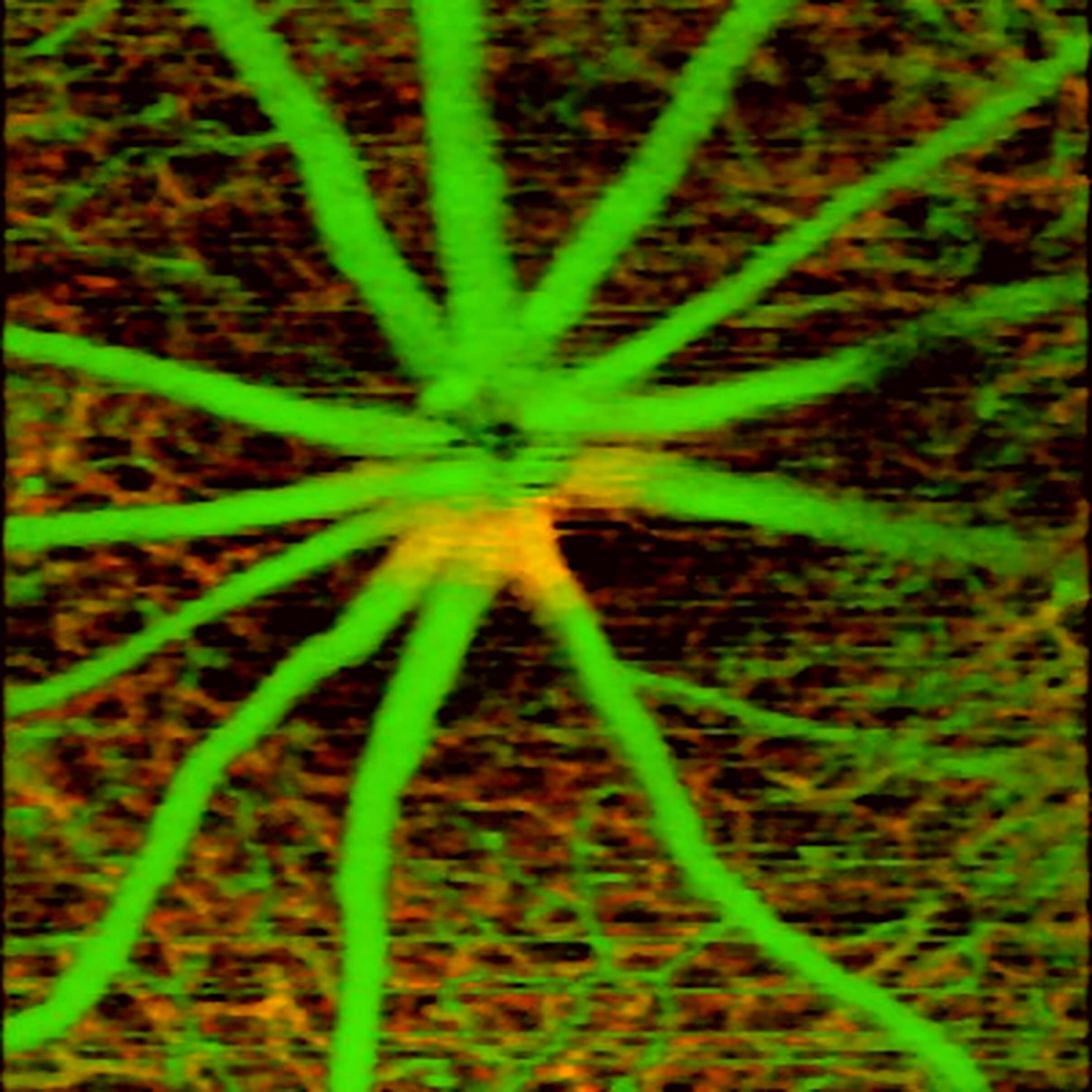
OCT angiography image of mice retina shows both large and micro-vessels at different depth locations using intrinsic contrast only. The false color in the image encodes the depth location of the blood vessels. Image taken with 800 nm system using a Cobra 800 spectrometer. The vasculature contrast was generated using OCT angiography.
Misty Johnson is responsible for taking care of our grating customers at the Logan, Utah, facility. She has been a member of the Wasatch Photonics team since 2009. By understanding her customers’ needs and goals, as well as the current grating market, Misty is able to provide a superior level of customer support. As Account Manager, she acts as a liaison between customers and Wasatch Photonics’ manufacturing department making sure that the lines of communication are always open.
Customer support is an integral part of Wasatch Photonics commitment to customers. Whether you are in the market to purchase a single grating, inquiring as a distributor, or purchasing large volume, Misty will guide you seamlessly through the custom design process. Misty strives to build trust and strong long term relationships with Wasatch Photonics customers. She brings 20 years’ experience as customer support manager from a number of manufacturing and service companies.