A rapid detection technique for subsurface, cross-sectional visualization of materials
Back in 1991, David Huang, Eric Swanson, James Fujimoto, and colleagues from MIT and Harvard Medical School published the first paper, in Science, on Optical Coherence Tomography (OCT). In fact, it introduced the term and showcased the ability to produce cross-sectional images of the human retina with micrometer-level resolution. As such OCT began in ophthalmology and it is in this field it continues to excel. In recent times, new application areas have opened, all relying on the non-invasive methodology for sub surface diagnostics.
What is OCT?
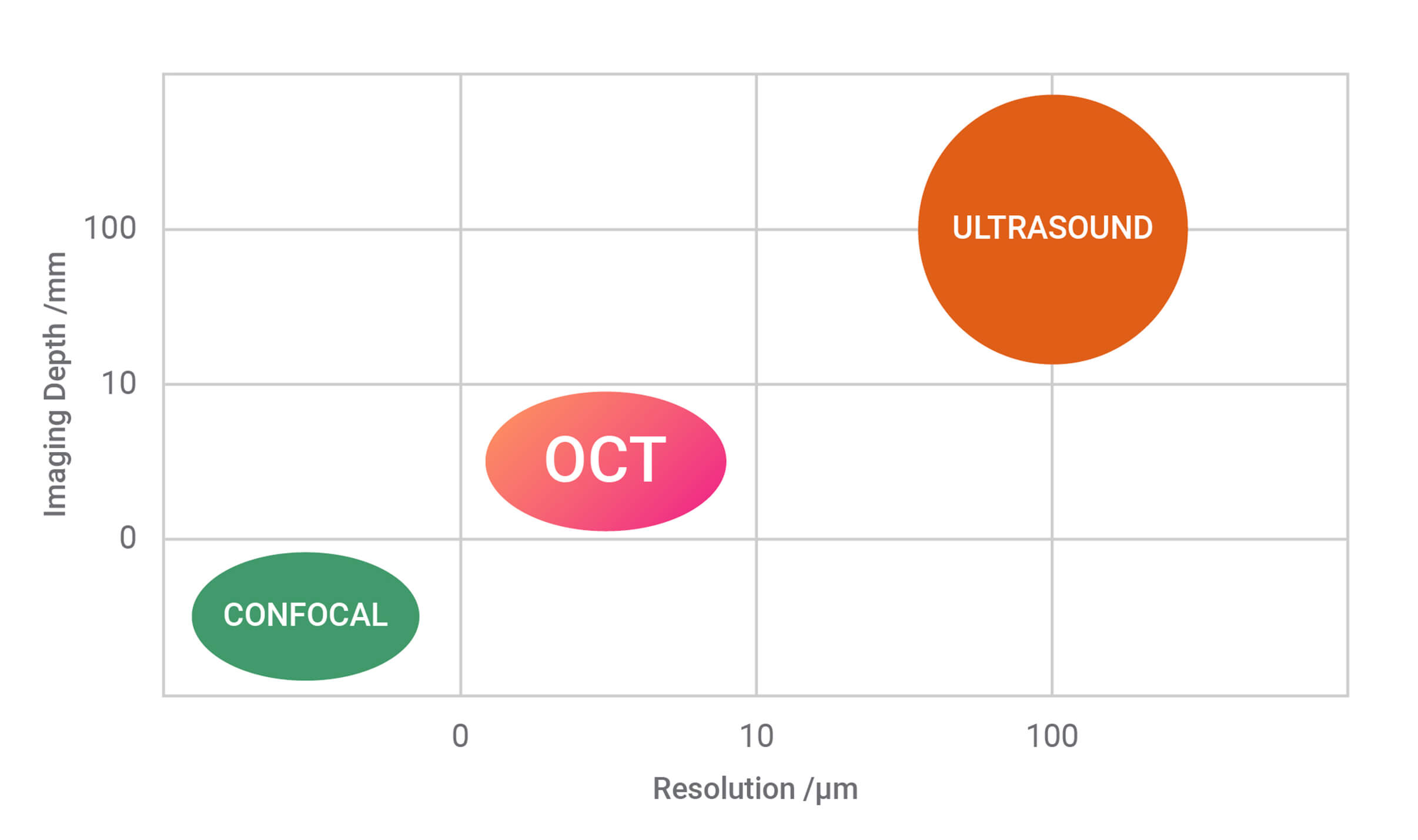
OCT is a 3-D imaging technique that can provide high resolution imaging in scattering media, non-destructively and without the need for contact or a coupling medium. Axial imaging resolution on the order of a few micrometers is possible down to depths of 14 millimeters. OCT’s ability to provide surface profiles and information about subsurface structure and uniformity enables it to deliver accurate information in real time for diagnostics, monitoring, and in-situ process feedback.
How Does OCT Work?
OCT relies on back-scattered light from different regions of a sample to generate a 3D image. It uses different localization techniques to obtain information in the axial direction (along the optical beam or into the sample, z-axis) and the transverse direction (plane perpendicular to the beam or across the sample, x-y axes). The information in the axial direction is obtained by measuring the time delay of light reflected from structures or layers in the sample. This technique is similar to that which is used to generate an ultrasound image, with light being used instead of sound. Given the high speed of light, it is not easy to perform a direct measurement of the time delay for the back-scattered light. Instead, OCT systems indirectly measure the time delay using what is called low-coherence interferometry.
Introducing Low Coherence Interferometry
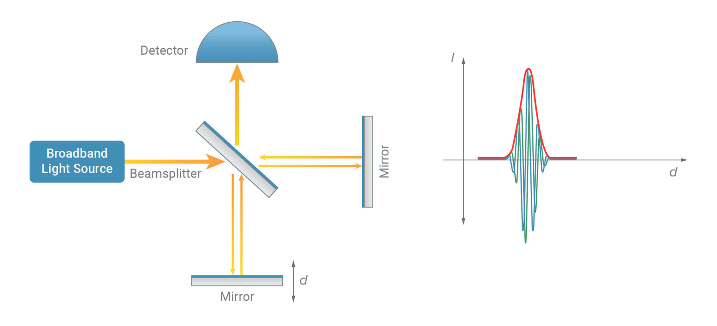
In a low-coherence interferometer, a light source with a broad optical bandwidth is used for illumination. The light coming out of the source is split by a beam splitter into two paths called the reference and sample arms of the interferometer. The light from each arm is reflected back and combined at the detector. An interference effect (fast modulations in intensity) are seen at the detector only if the time travelled by light in the reference and sample arms is nearly equal. Thus, the presence of interference serves as a relative measure of distance travelled by light.
Low-coherence interferometry at the concept level. The mirror arm is kept fixed, and interference between the two arms is measured.
Introducing Optical Coherence Tomography
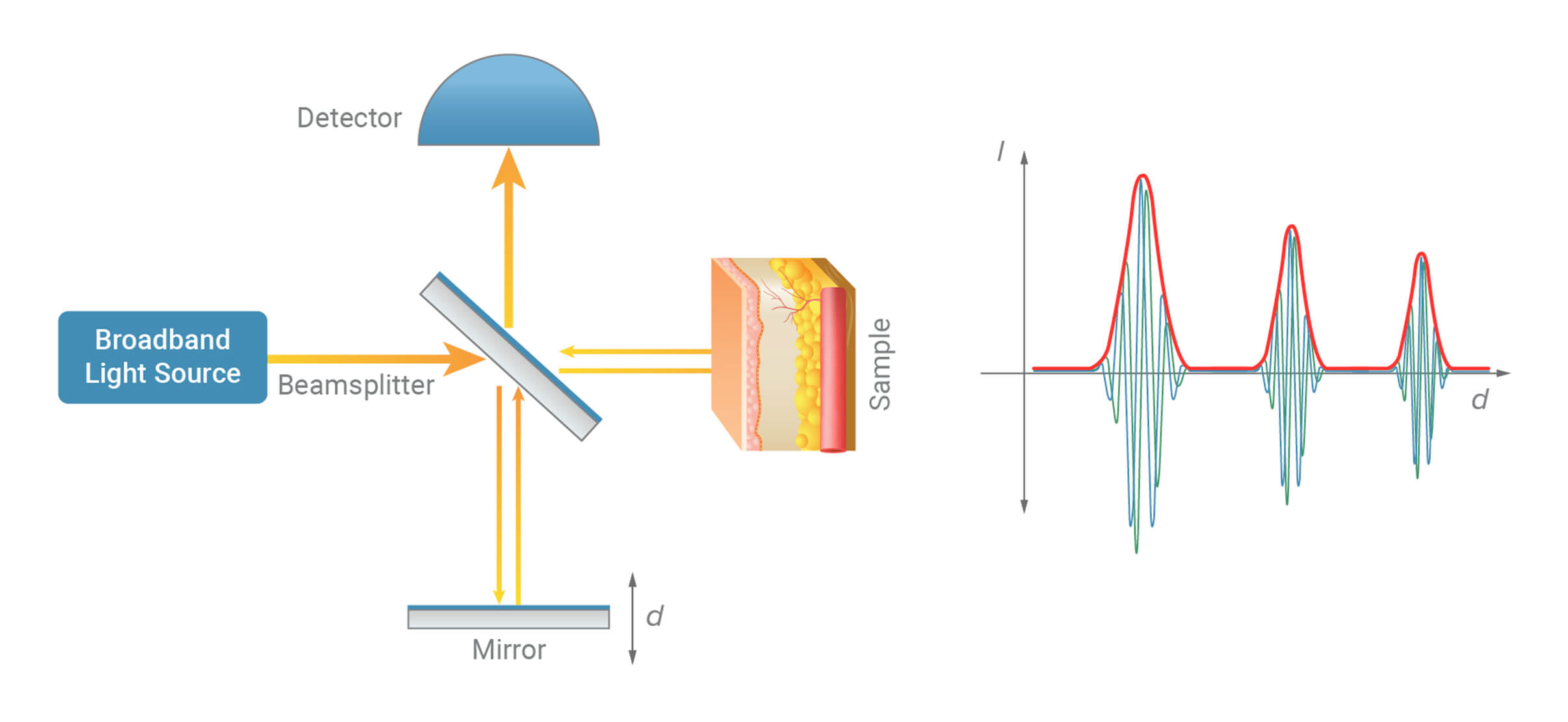
Optical coherence tomography (OCT) builds on this concept by replacing the mirror in the sample arm with the sample itself. The reference arm is scanned in a controlled manner while the resulting light intensity is recorded by the detector. Interference occurs when the optical path length of the reference mirror matches that of a reflecting structure within the sample. This interference signal can then be processed to identify the presence and location of that structure.
The distance between two reference mirror positions that produce interference corresponds to the optical distance between two reflecting structures within the sample along the beam path. Although the light beam interacts with multiple structures as it travels through the sample, low-coherence interferometry allows the reflections from each structure to be distinguished. In this way, OCT measures the scattering properties of the material as a function of depth, enabling depth-resolved structural imaging.
Introducing Fourier-Domain OCT
Fourier-domain OCT allows for much faster imaging than scanning of the sample arm mirror in the interferometer, as all the back reflections from the sample are being measured simultaneously. This speed increment introduced by Fourier-domain OCT has opened a whole new arena of applications for OCT.
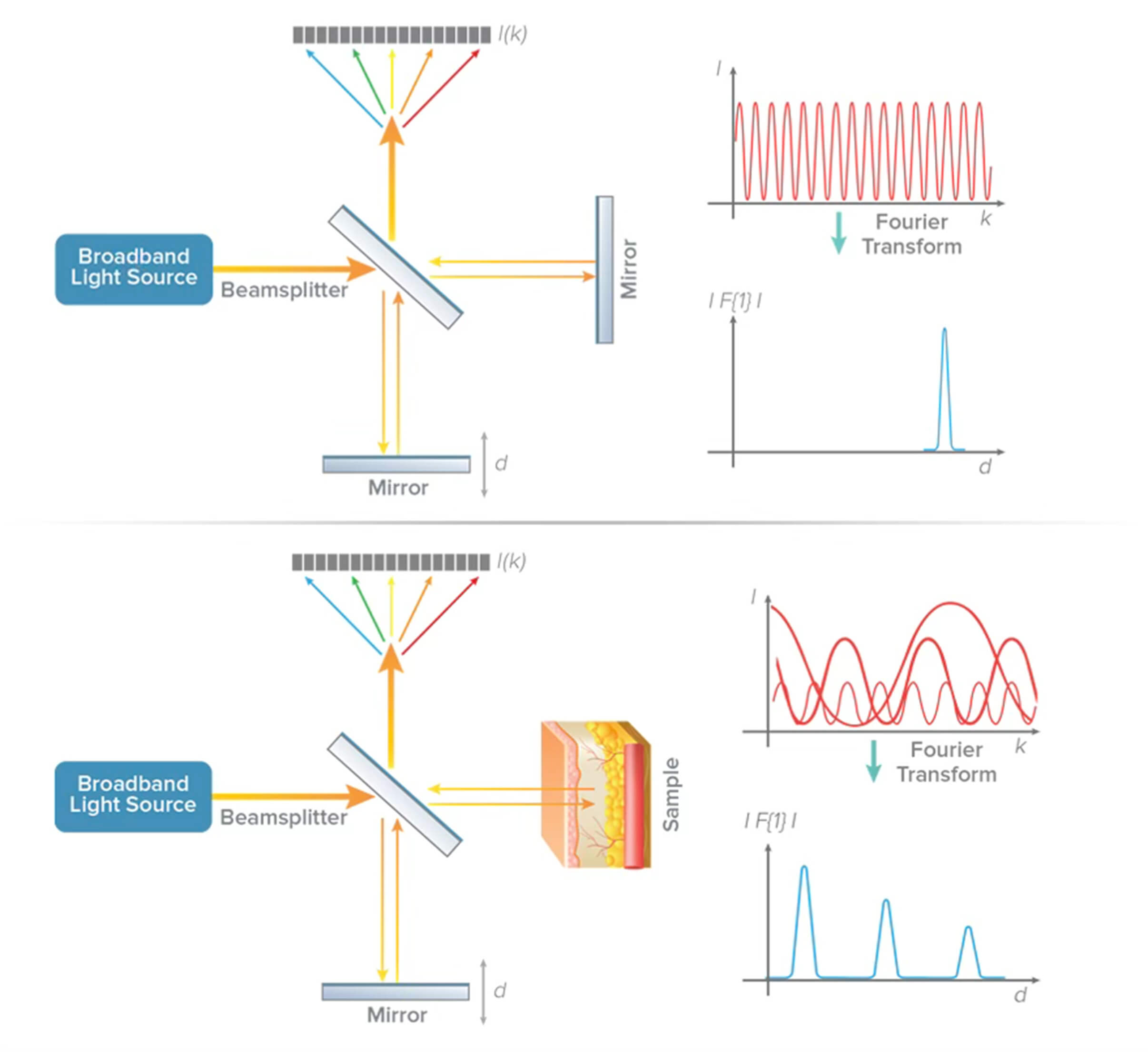
Fourier-domain OCT (FD-OCT) provides an even more efficient way to implement the low-coherence interferometry. Instead of recording intensity at different locations of the reference mirror, the intensity is recorded as a function of the wavelengths or frequencies of the light. The intensity modulations when measured as a function of frequency are called spectral interference. The rate of variation of intensity over different frequencies is indicative of the location of the different layers reflected in the samples. The Fourier transformation of this spectral interference data provides information equivalent to that which would be obtained by moving the reference mirror.
Fourier-domain OCT, in which the mirror arm is kept fixed and interference as a function of wavelength is recorded.
Introducing Spectral-Domain OCT
There are two common methods of measuring spectral interference in OCT: spectral-domain and swept-source. In spectral-domain OCT (SD-OCT), a broadband light source delivers many wavelengths to the sample, and all are measured simultaneously using a spectrometer as the detector. In swept-source OCT (SS-OCT), the light source is swept through a range of wavelengths, and the temporal output of the detector is converted to spectral interference.
Misty Johnson is responsible for taking care of our grating customers at the Logan, Utah, facility. She has been a member of the Wasatch Photonics team since 2009. By understanding her customers’ needs and goals, as well as the current grating market, Misty is able to provide a superior level of customer support. As Account Manager, she acts as a liaison between customers and Wasatch Photonics’ manufacturing department making sure that the lines of communication are always open.
Customer support is an integral part of Wasatch Photonics commitment to customers. Whether you are in the market to purchase a single grating, inquiring as a distributor, or purchasing large volume, Misty will guide you seamlessly through the custom design process. Misty strives to build trust and strong long term relationships with Wasatch Photonics customers. She brings 20 years’ experience as customer support manager from a number of manufacturing and service companies.