Superior depth & contrast for highly scattering samples
Visualization of deep-tissue structures can be limited in penetration depth and contrast, particularly for samples with high scattering power & low water content, such as lipid-rich tissue, dental structures, and even paints and rubber.
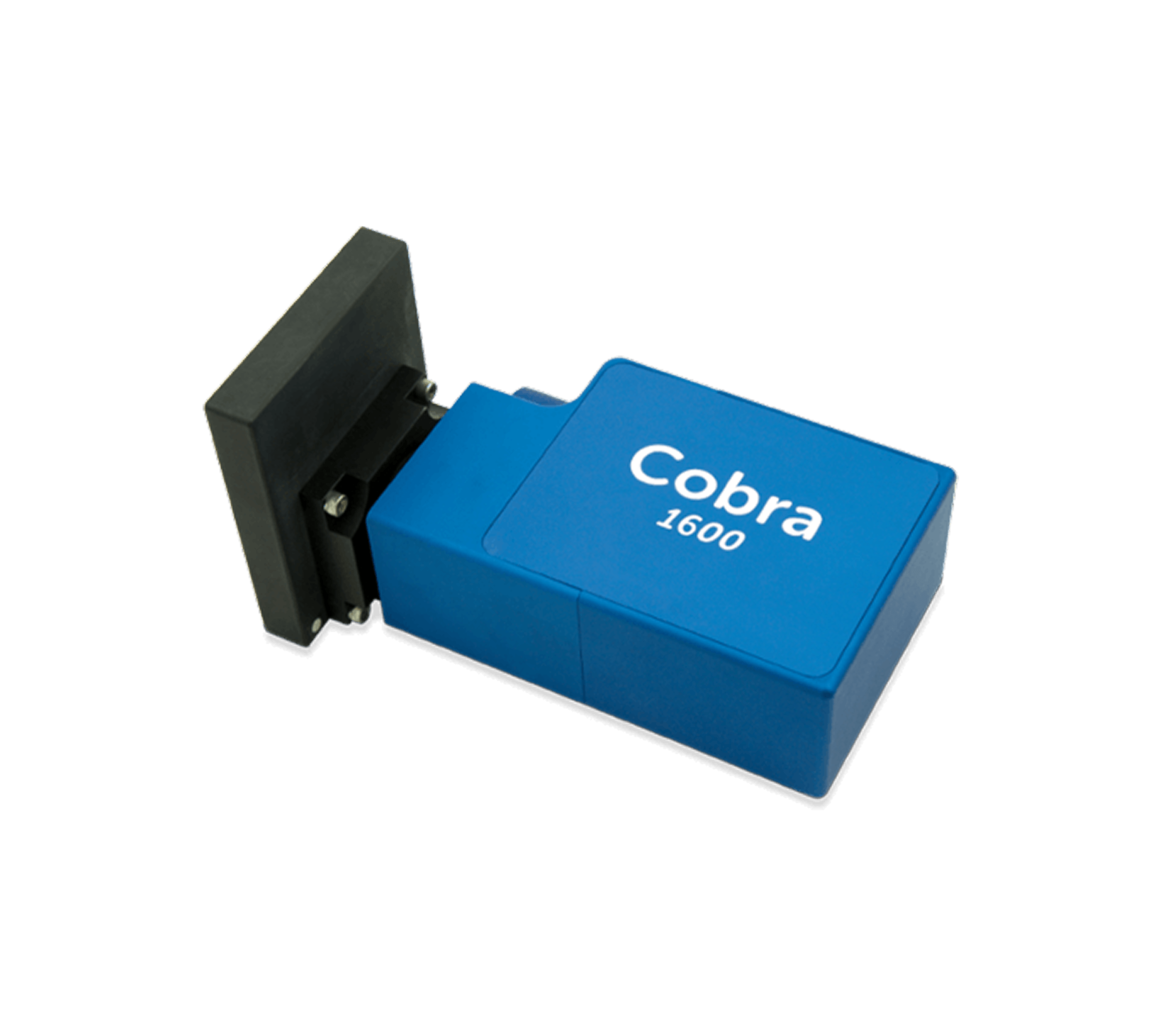
As the first commercially available off-the-shelf 1600 nm OCT spectrometer, the Cobra 1600 gives you access to an optical window with enhanced depth penetration and high contrast with high axial resolution, compatible with readily available fiberoptic components. Our proprietary high throughput design offers the sensitivity & speed needed for 3D deep-tissue imaging to study hippocampal formation and characterize atherosclerotic plaques, or for non-destructive testing.
Cobra 1600 OCT spectrometers can be used for en face and 3D imaging in research, industrial and OEM solutions.
240 nm bandwidth for high resolution at long wavelength
•
Premier optical design with superior SNR & subpixel resolution for greater image clarity
•
Near diffraction-limited optics minimize roll-off
•
Proprietary VPH gratings for low polarization dependence & highest efficiency
•
Ability to image 25% deeper in highly scattering tissues than 1300 nm OCT
•
OEM ready: robust & compact
Spectrometer Models
C1600-1570/240-147-SG2K
Wavelength Range
1450–1690 nm
Bandwidth
240 nm
Imaging Depth
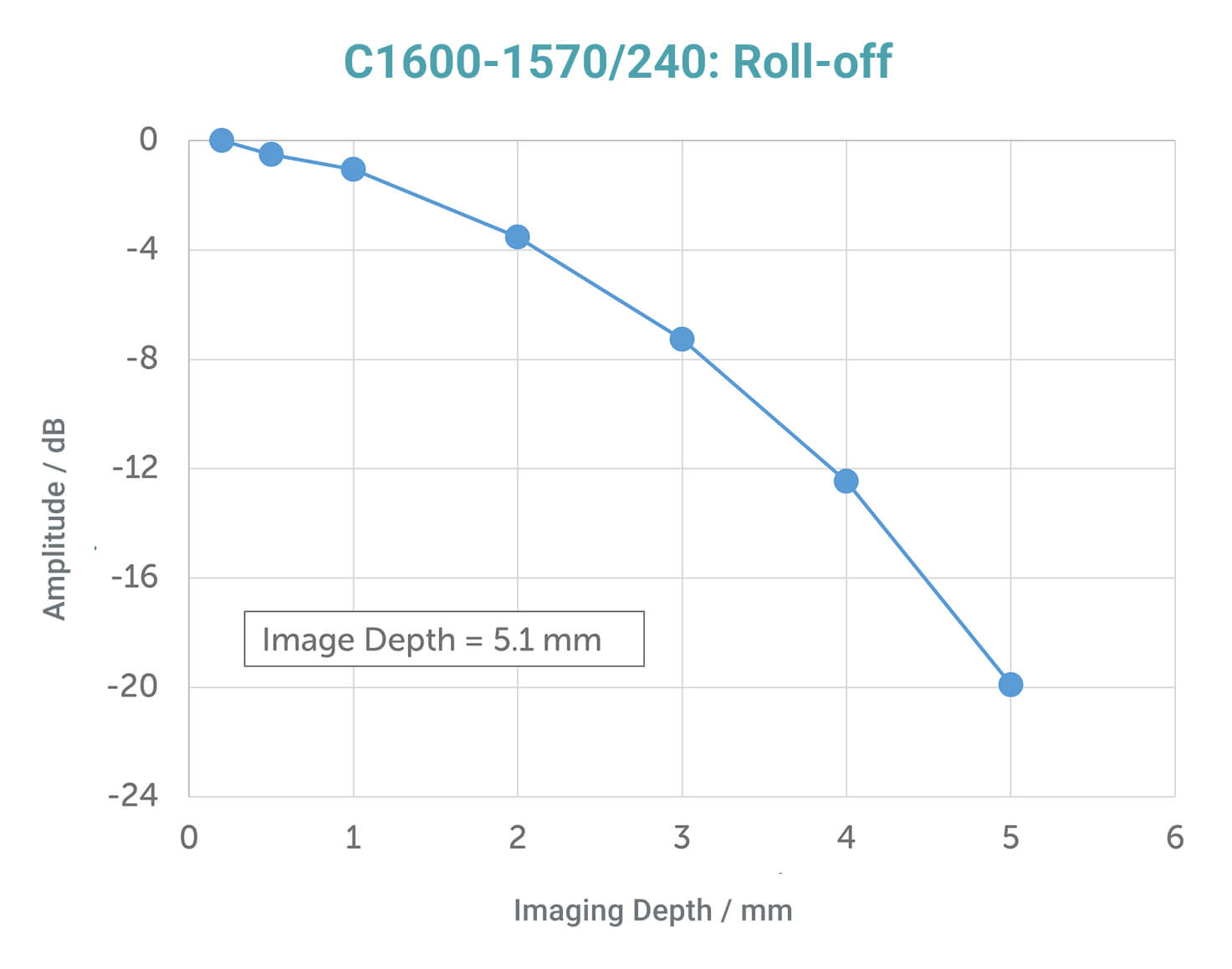
5.1 mm
Spectral Resolution
0.12 nm
Axial Resolution
4.5 µm
Max Line Rate
147 kHz
Pixels
2048
Interface
Camera Link
Dimensions
9.5 cm x 19 cm x 6 cm
Weight
1.4 kg
Cobra 1600 part numbers are of the format C1600-[CWL]/[BW]-[kHz]-[camera][pixels], where the Sensors Unlimited GL2048 camera is represented by “SG”, and 2K denotes 2048 pixels. Imaging depth stated is the theoretical value, calculated in air.
Camera Models
•
Camera Link: 147kHz
Roll-off Performance
The Cobra 1600 OCT spectrometer offers superior roll-off performance, as can be seen in this comparison of signal with imaging depth.
Applications
•
Optical coherence microscopy
•
Intravascular OCT
•
Non-destructive analysis
SDKs & Software Support
Compatible with Wasatch Photonics’ SDKs for rapid data access in development environments (e.g., C++, LabVIEW, MATLAB).
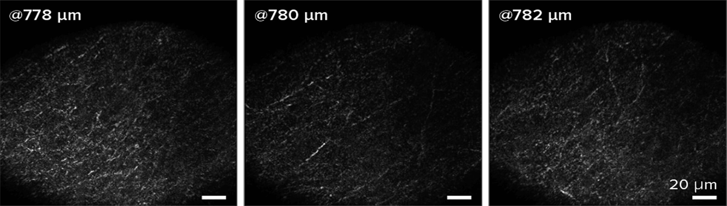
In Action
Optical coherence microscopy imaging of mouse brain in vivo. OCM en face images were taken at 3 successive depths, with imaging NA = 1. Courtesy of Steven Adie, Cornell University.